#PhDone: A molecular dissection of vomeronasal neurons
Most of us have seen dogs sniff rear ends. By doing so, they are trying to detect chemical molecules to determine identity, sex and menstrual state of other dogs. Most animals communicate with each other, and the outside world, by sensing chemical molecules using their olfactory system.

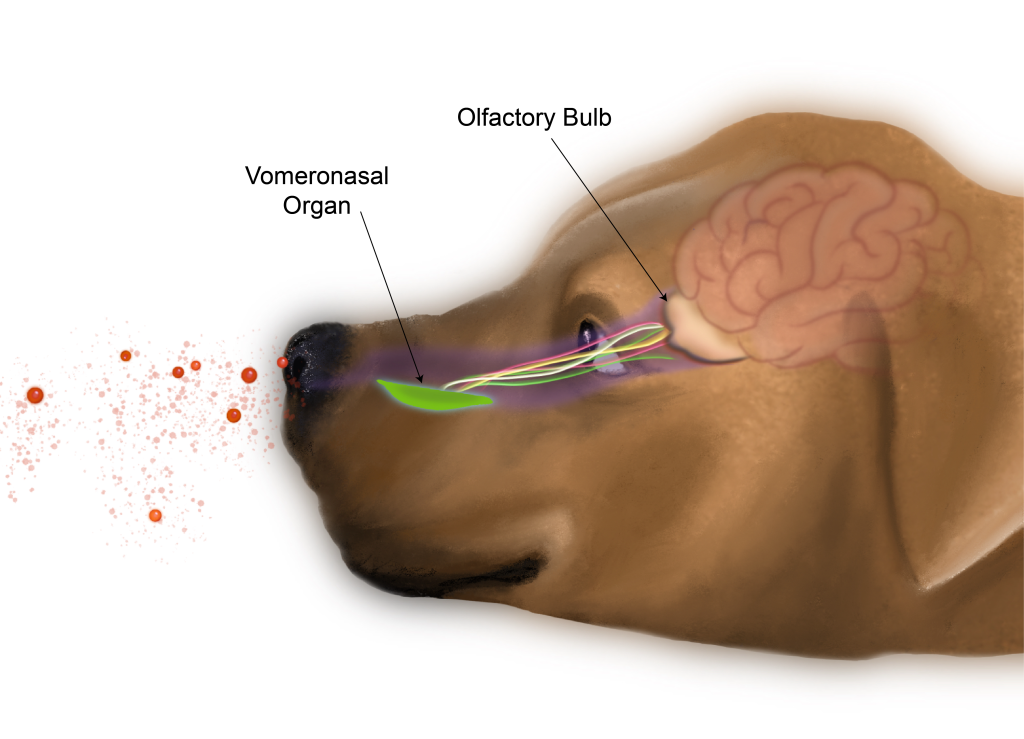
Illustration: Soumita Samanta
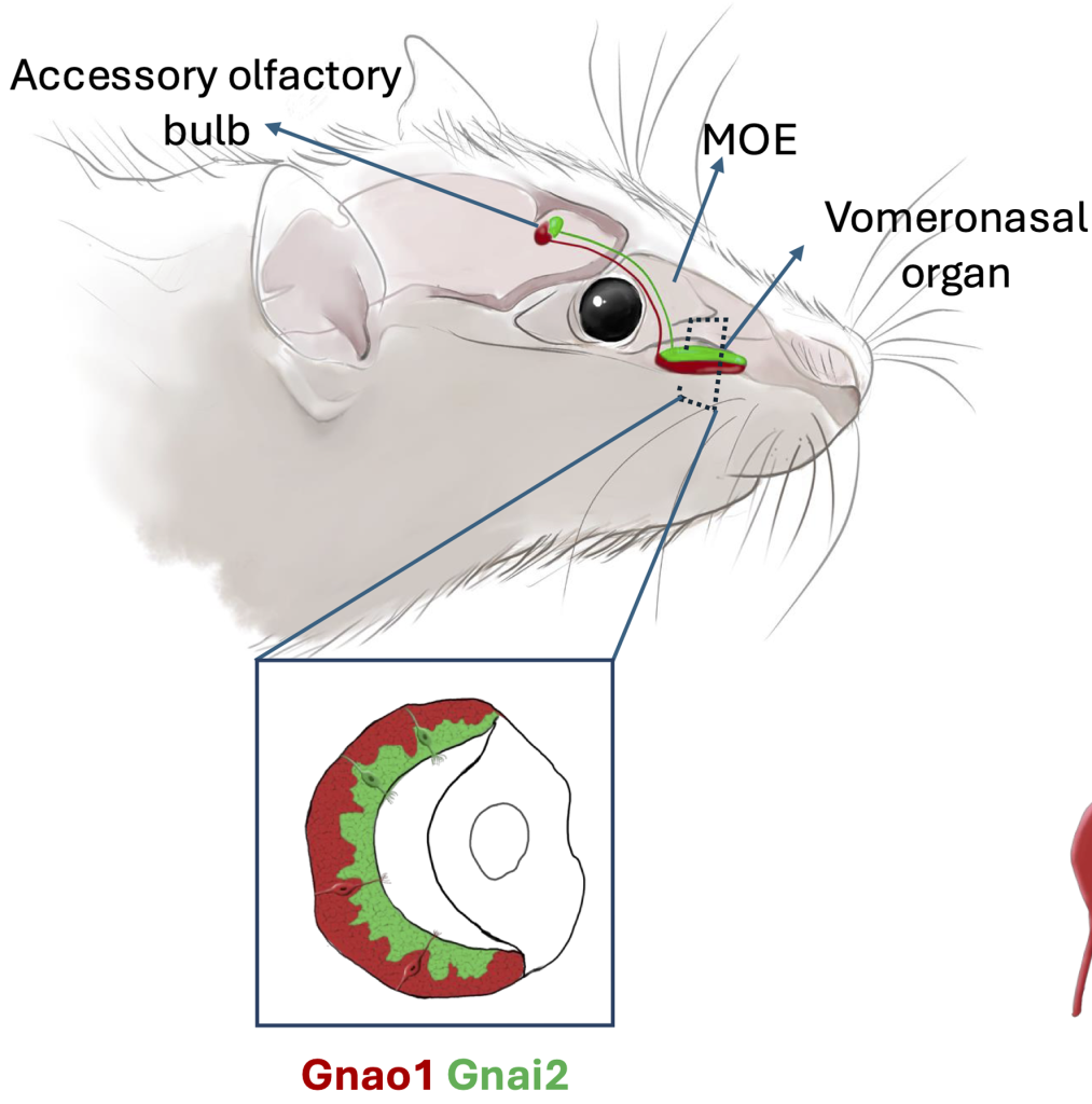
A) The vomeronasal organ (VNO), a part of the accessory olfactory system, is responsible for detecting odorants, mostly pheromones. The neurons in the VNO are known to develop from a common stem cell population and diversify during development into two broad cell types – Gnao1 and Gnai2. These express two different families of pheromone receptors.
Using mice as a model system, Devakinandan probes gene expression profiles in these two neuron subtypes. His major finding is that in addition to sensory receptors, the two cell types differ in endoplasmic reticulum (ER) structure, content and gene expression. Further he identifies novel ER-resident proteins that are unique to these neurons.
Image: GVS Devakinandan, PhD Thesis
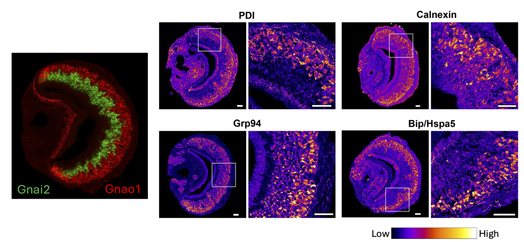
Differential expression of ER proteins – PDI, Calnexin, Grp94, Hspa5 in Gnao1 and Gnai2 neurons. Image: GVS Devakinandan, et al. eLife 13 (2024): RP98250
B) To compare gene expression between the two major sensory neuron subtypes Devakinandan used the single cell transcriptomics data and found that one of the subtypes selectively upregulated expression of genes that are related to endoplasmic reticulum (ER) functions.
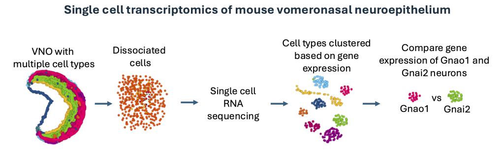
Image: GVS Devakinandan, PhD Thesis
C)
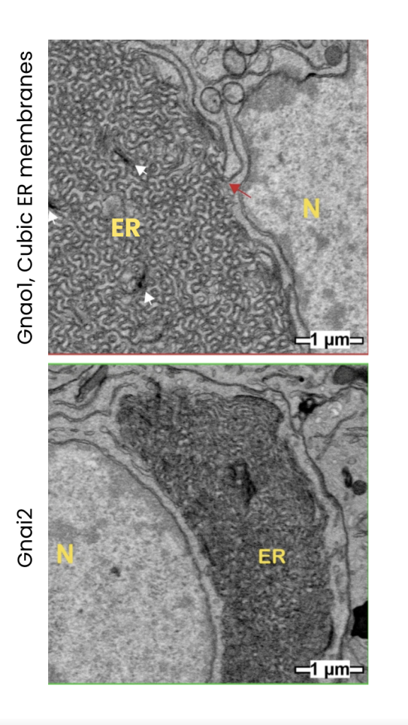
Electron microscopy studies reveal that Gnao1 neurons are densely packed ER membranes that adopt a gyroid (cubic) ultrastructure.
Image: GVS Devakinandan et al. eLife 13 (2024): RP98250. (N= Nucleus, ER= Endoplasmic Reticulum)
This study raises an intriguing question about how the ER may contribute to the development of sensory neurons and consequently enable their function. These findings take us a step closer towards gaining a better understanding of animal olfaction and pheromonal communication at the molecular level.
Read more about this work here:
– Single-cell transcriptomics of vomeronasal neuroepithelium reveals a differential endoplasmic reticulum environment amongst neuronal subtypes.